Babyecho for Dummies
Babyecho for Dummies
Blog Article
What Does Babyecho Do?
Table of ContentsNot known Details About Babyecho 9 Easy Facts About Babyecho ShownAn Unbiased View of BabyechoLittle Known Questions About Babyecho.Babyecho for BeginnersGetting My Babyecho To WorkSome Known Details About Babyecho
:max_bytes(150000):strip_icc()/JoseLuisPelaezInc-17f79a53211940c2bc62cf23bc4185d4.jpg)
A c-section is surgical treatment in which your child is birthed with a cut that your physician makes in your stubborn belly and uterus. No matter what an ultrasound reveals, speak to your company about the most effective take care of you and your infant - at home doppler. Last assessed: October, 2019
Throughout this scan, they will examine the baby is growing in the best area, whether there is greater than one infant and they will likewise examine your infant's advancement up until now. This testing is available in between 10 14 weeks of maternity and is made use of to evaluate the possibilities of your child being born with several of these conditions.
Everything about Babyecho
It includes a mixed test of an ultrasound check and a blood test. During the scan, the sonographer will determine the liquid at the rear of the infant's neck to identify 'nuchal clarity' - https://pblc.me/pub/3cb9f10e6009b1. They will then calculate the chance of your baby having Down's, Edwards' or Patau's disorder utilizing your age, the blood examination and check outcomes
Throughout this scan, the sonographer checks for architectural and developing problems in the baby. Throughout this check visit, you may be offered screenings for HIV, syphilis and liver disease B by a professional midwife. In many cases, a third-trimester scan is suggested by your midwife complying with the results of previous tests, previous difficulties or existing medical conditions.
The traditional 2D ultrasound generates flat and detailed pictures which can be used to see your child's interior organs and assist discover any inner concerns. These black and white images assist the sonographer identify the child's pregnancy, development, heartbeat, development and dimension. Some expectant mothers pick to have a 3D ultrasound scan due to the fact that they reveal more of a real-life picture of the infant.
Getting The Babyecho To Work
3D ultrasound scans reveal still pictures of your infant's exterior body instead of my sources their withins, so you can see the shape of the child's face functions. 4D ultrasound scans resemble 3D scans however they reveal a moving video clip as opposed to still pictures. This catches highlights and darkness better, for that reason developing a clearer photo of the baby's face and motions.

or (8-11 weeks) (11-14 weeks) (14-18 weeks) (19-23 weeks) or (24-42 weeks) Advised at Optional -, extra frequently in some problems This check is done to and to determine an (EDD). A is identified throughout this check. A lot of parents go with this check for. Likewise is important prior to the blood test called as (NIPT) to calculate the.
Our Babyecho Ideas
Occasionally a might be required to get and a clearer image. This is typically executed and sometimes a may be required (baby heart monitor). Validate that the child's heart is existing; To much more precisely.
Please see below. These scans might be done, nevertheless some of the and hence, a is required to This check is done generally at.
Facts About Babyecho Uncovered

Furthermore, the can be by by an. () The means nearer the is helpful to. Sometimes, an which was previously may be.
6 Simple Techniques For Babyecho
If, these scans might be to. on the searchings for, a may be provided. During all the, a 3D check (of the child) can additionally be performed. The depends on the position of the,,, quantity of and. This includes, together with; This includes, together with (14-20 weeks).
A check is vital before this test is done. If you're seeking, arrange a consultation with Dr Sankaran via her. Obstetrics & gynaecology in London.
How Babyecho can Save You Time, Stress, and Money.
The examination can offer valuable details, assisting females and their health-care companies take care of and care for the pregnancy and the unborn child.
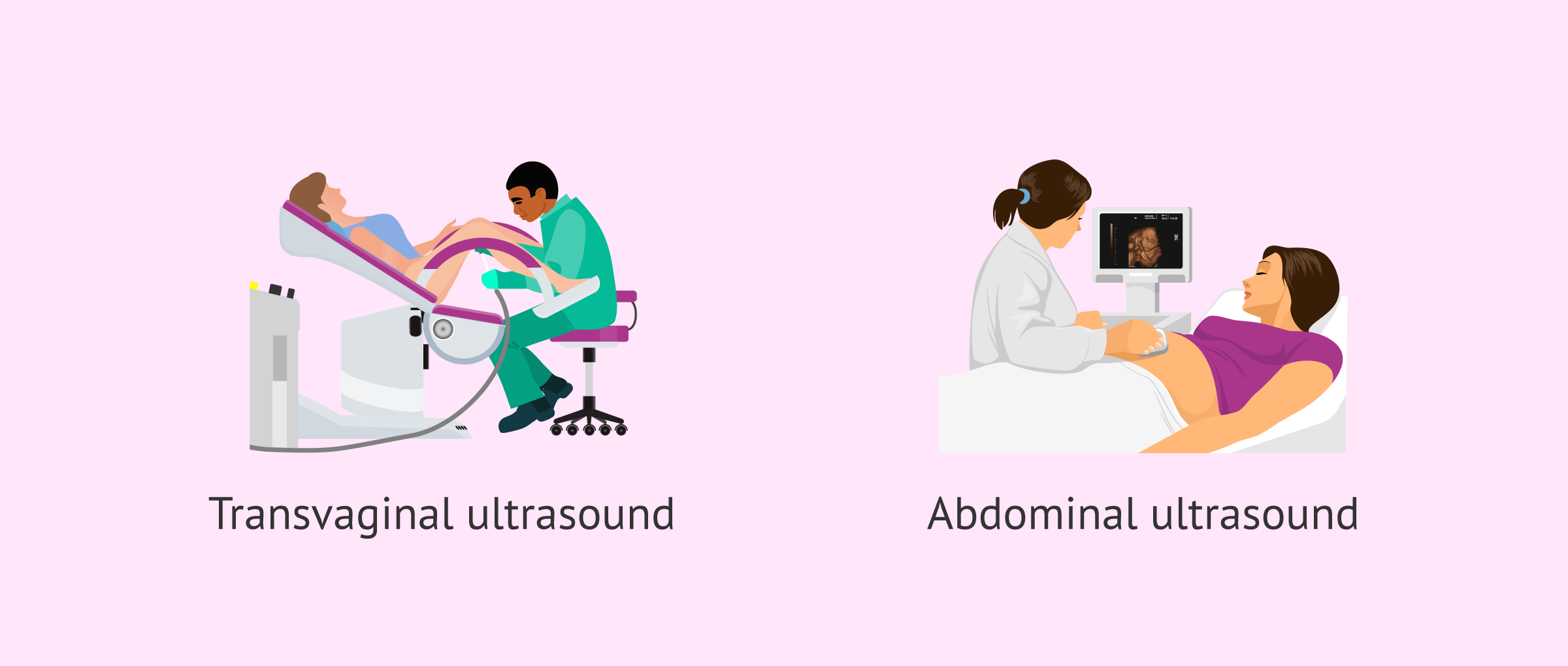
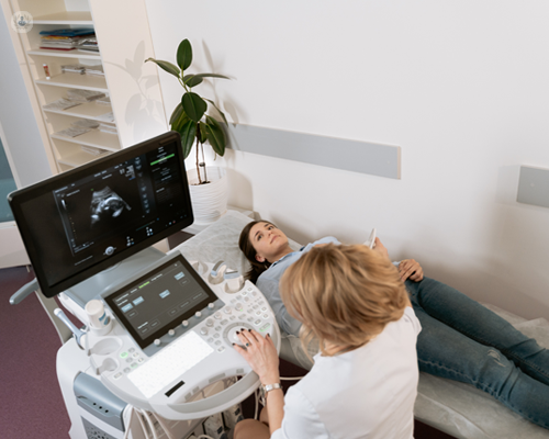
A transducer is placed into the vaginal area and rests against the back of the vaginal canal to develop a photo. A transvaginal ultrasound produces a sharper picture and is often utilized in early pregnancy. Ultrasound devices have to do with the dimension of a grocery cart. A television screen for seeing the photos is attached to the equipment (https://www.brownbook.net/business/52713786/babyecho/).
Report this page